Factors that contribute to this include poor sensitivity of the history and physical exam, a broad differential diagnosis that crosses several specialties, and an often negative diagnostic workup. [4]Sandler RS, Stewart WF, Liberman JN, et al. Although sonography is the preferred method for diagnosis of acute cholecystitis, CT is frequently the initial examination because the diagnosis is unclear. AL has been the principal applicant/co-applicant for a number of grants including: Engineering and Physical Sciences Research Council Research - Multiplexed AKI biomarker detection with a single molecule biosensor; Leeds Cares - A novel, non-invasive diagnostic approach to assess kidney transplant health through the targeted measurement of biomarkers of kidney injury and immune response in kidney transplant recipients at the Leeds Teaching Hospitals NHS Trust; Kidney Research Yorkshire - Use of enhanced technology to characterise haemodialysis treatment for acute kidney injury (AKI); Bringing It Home - Validation of a micro-sampling technique for measuring tacrolimus and creatinine remotely; and to fund a research nurse; British Renal Society - Renal function assessment with point of care creatinine in diverse populations (RAPID), and several NIHR grants including on a) Improving the quality of post-discharge care following AKI b) An investigation into the use of remote blood sample collection to reduce health inequalities in patients with mental health disorders c) A comparison of remote blood collection devices: a human factor use study d) Defining the characteristics for a novel automatic device to monitor urine output in catheterized patients e) Leeds Medtech and In-vitro Diagnostic Cooperative Grant Extension f) Surgical MedTech Co-operative for the 2019/20 proof-of-principle funding stream g) A pilot investigation into the use of beta-trace protein for residual renal function estimation in haemodialysis h) Application of functional MRI to improve assessment of chronic kidney disease (AFiRM study) i) SuperResPath-Renal: Quantitative super-resolution technology for a fast, decentralised clinical diagnosis of renal pathologies. Tests and procedures used to diagnose A suspected diagnosis of acute cholecystitis is best confirmed by ultrasonography, with the visualization of gallstones in the gallbladder. Often, the cause of peritonitis can be detected only intraoperatively. For any urgent enquiries please contact our customer services team who are ready to help with any problems. PLoS One. These digestive enzymes are secreted in their inactive form and are activated by an enzyme once in the duodenum. Diagnosis consists in conducting a surgical examination, laboratory tests, ultrasound, X-ray examination and CT of the abdominal cavity. Listed below are three potential differential diagnoses and their relation to the patient presentation provided on the previous page. Please try after some time. Acute cholecystitis develops in up to 10 percent of patients with symptomatic gallstones and is caused by the complete obstruction of the cystic duct. Abdominal pain is often differentiated with acute appendicitis, cholecystitis, right-sided paranephritis and cholelithiasis. FA declares that he has no competing interests. The mucosa may be simplified and relatively atrophic secondary to healing of erosions. 28-12). JC declares that she has no competing interests. Am J Epidemiol. When the gallbladder is filled sufficiently with excreted tracer (typically 30-60 minutes after tracer injection), sincalide is then administered. In cases of chronic cholecystitis, these results may be within the reference range. There can also be damaged to the acinar cells due to an obstruction of flow such as from a gallstone, which creates inflammation and edema. Thorotrast agent: Used in medical imaging until 1960; long latency for development of cancer of 20 to 40 years. MC declares that none of these interests is directly related to topics advised on for this project. The gallbladder may be normal, distended or thickened. Pathophysiology & Clinical Presentation Correct Diagnosis. 2007 Jun;56(6):815-20. 28-13). Neither text, nor links to other websites, is reviewed or endorsed by The Ohio State University. Dig Dis Sci. Diagnosis and management of patients with chronic abdominal pain is often challenging, and can be a frustrating experience for both physicians and patients. Figure 4. [25] A combination of Ultrasound is the definitive initial test. On ERCP, a normal intra- and extra-hepatic biliary duct; there was CBD sludge but no CBD stones. AA sits on advisory committees for the following research studies: VOICE2 (development and testing of communication skills training for hospital healthcare practitioners caring for people living with dementia), SWOP (social work in older people) and CFIN (cognitive frailty interdisciplinary network). Gastroenterology. Patients with chronic cholecystitis present with symptoms of recurrent biliary colic. WebThere were 82 men and 49 women in the acute cholecystitis group (n = 131) and 107 men and 144 women in the chronic cholecystitis group (n = 251) (Fig. As such, patient medications should be reviewed at the time of the study. http://www.ncbi.nlm.nih.gov/pubmed/14625840?tool=bestpractice.com Chronic cholecystitis is a chronic condition caused by ongoing inflammation of the gallbladder resulting in mechanical or physiological dysfunction its emptying. 2007 Oct;61(10):1663-70. http://www.ncbi.nlm.nih.gov/pubmed/17681003?tool=bestpractice.com. In Nuclear Medicine (Fourth Edition), 2014. RUQ tenderness, a distended RUQ mass, and positive Murphy sign are key examination findings. GR has been paid for advisory board meetings with the following companies: Safoni Aventis, Abbott Diabetes UK, Lilly Diabetes, Bayer. to maintaining your privacy and will not share your personal information without
It has been suggested that chronic cholecystitis develops as a result of recurrent attacks of mild acute cholecystitis. http://www.ncbi.nlm.nih.gov/pubmed/10565617?tool=bestpractice.com CT abdomen with contrast showed thickening of the gall bladder wall. [6]Talley NJ, Weaver AL, Zinsmeister AR, et al. Magnetic resonance cholangiopancreatography may be required. Magnetic resonance cholangiopancreatography or computed tomography are both useful tools in the visualization of the common bile duct and pancreatic ducts and can detect stones in both the common bile duct and in some cases stones near the ampulla. Scand J Gastroenterol Suppl. -, Benkhadoura M, Elshaikhy A, Eldruki S, Elfaedy O. 2018 Dec;121:131-136. The PubMed wordmark and PubMed logo are registered trademarks of the U.S. Department of Health and Human Services (HHS). MC is chair of the European Society of Cardiology Digital Health Committee and a trustee of the Atrial Fibrillation Society. They will usually order an ultrasound of your abdomen. the term of Cholecystitis it is referred to inflammation in the gallbladder and it is considered as a surgical condition. 2015 Mar;12(3):159-71. The differential diagnosis of xanthomatous cholecystitis includes mycobacterial and fungal infections, which generally result in better-formed granulomas and are excluded by use of special stains. In some cases, the histiocytes contain brown ceroid pigment. Organic etiologies have a clear anatomic, physiologic, or metabolic cause. Clinical manifestations develop rapidly: there is acute intense pain in the right hypochondrium, vomiting, bloating, hypotension and tachycardia, symptoms of intoxication increase. [1], Associate Editor(s)-in-Chief: Furqan M M. M.B.B.S[2]. ASB declares that he has no competing interests. Treasure Island (FL): StatPearls Publishing; 2023 Jan. Despite the rapid development of surgery, mortality remains high, ranging from 20 to 35%, depending on the causes of peritonitis. Chronic intestinal pseudo-obstruction: Etiology, clinical manifestations, and diagnosis; Clinical manifestations and diagnosis of early pregnancy; Clinical manifestations and diagnosis of gastroesophageal reflux in adults; Cyclic vomiting syndrome; Eating disorders: Overview of epidemiology, clinical features, and diagnosis 2023. In an emergency, surgical intervention is performed to eliminate the source of peritonitis, antibiotics, painkillers and antishock drugs, parenteral solutions are prescribed. [3]Heading RC. Epidemiologic studies suggest that the vast majority of patients with chronic abdominal pain have functional GI disorders such as irritable bowel syndrome or functional dyspepsia. This occurs from injury to the acinar cells, which allows the digestive enzymes to be released and activated while still inside of the pancreas. Although the exact pathophysiology for appendicitis is unknown a common theory is that the lumen becomes obstructed. Chronic cholecystitis must also be differentiated from colitis, functional bowel syndrome, hiatal hernia, and peptic ulcer disease. A physical exam with a comprehensive history is paramount in making the diagnosis of cholecystitis. An official website of the United States government. Because many patients with chronic cholecystitis have an antecedent history of an episode of acute cholecystitis, children may be less likely to present with this particular entity. Based on the prevalence of the pathological process, there are: The clinical picture of the disease depends on the rate of penetration and the amount of bile entering the abdominal cavity, the area of the lesion. Further confusing this picture, serum cancer antigen 19-9 levels can be elevated in xanthogranulomatous cholecystitis (Clarke etal, 2007). Because chronic cholecystitis is almost always associated with cholelithiasis, the demographic characteristics of these patients and risk factors are the same as for cholesterol gallstones. In a patient with suspected sepsis, use computed tomography (or magnetic resonance imaging)to identify the cause. At all stages of the disease, emergency surgical intervention is carried out, aimed at getting rid of the pathology that led to the development of peritonitis (perforation of the gallbladder, failure of surgical sutures, etc.). The site is secure. This new onset of pain that is not resolving would indicate that this may not be a duodenal ulcer and Murpheys sign would not be present. In most cases (90%), it is caused by complete cystic duct obstruction usually due to an impacted gallstone in the gallbladder neck or cystic duct, which leads to inflammation within the gallbladder wall. Keefer L, Drossman DA, Guthrie E, et al. [25] A combination of 2 or 3 of the 4 CT findings could provide diagnosis and differentiation of acute cholecystitis from chronic cholecystitis with appropriate confidence. When present, the inflammatory pattern in patients with chronic acalculous cholecystitis is nonspecific. A clear relationship with an anatomic structure or underlying process may not always be present. Treatment of all types of cholecystitis is cholecystectomy as 90% of patients become asymptomatic. Centrally mediated disorders of gastrointestinal pain. 2005;15(3):329-38. doi: 10.1615/jlongtermeffmedimplants.v15.i3.90. 2000 Jun;45(6):1166-71. The surface epithelium may show regenerative hyperplasia but is more frequently denuded. Onset and disappearance of gastrointestinal symptoms and functional gastrointestinal disorders. About 1213% of patients with chronic cholecystitis do not have gallstones.103 It has been suggested that post-inflammatory stenosis or anatomic abnormalities of the cystic duct might impede normal emptying of the gallbladder. Imperial College London (Royal Brompton Hospital). Histologically, they comprise an admixture of fibrosis, myoid cells and glandular elements, thus the name fibromyoglandular polyps192 (Fig. The disease refers to life-threatening conditions, occurs in the practice of doctors of various specialties: gastroenterologists, abdominal surgeons, resuscitators. 14.51). On microscopic examination, large numbers of histiocytes with foamy cytoplasm admixed with chronic inflammatory cells are found. [2]Wallander MA, Johansson S, Ruigomez A, et al. Dysplasia is present in about 15% of cases, usually resulting from colonization by dysplasia and carcinoma involving the background mucosa. However, the pain associated with these disorders is nonspecific and can resemble or coexist with organic disorders. NS declares that he has no competing interests. WebThe article contains a description of various clinical "masks" of chronic cholecystitis, which make the diagnosis more difficult: cardial, duodenal (gastrointestinal), rheumatic, The etiology of chronic abdominal pain is so wide that only the more common causes can be covered here. 2003 Oct;33(4):279-96. Get new journal Tables of Contents sent right to your email inbox, Clinical and Translational Gastroenterology, Articles in PubMed by Rukevwe Ehwarieme, MD, Articles in Google Scholar by Rukevwe Ehwarieme, MD, Other articles in this journal by Rukevwe Ehwarieme, MD, Privacy Policy (Updated December 15, 2022). Acute Severe Pancreatitis. Peptic ulcers can manifest in many ways including single versus multiple and acute versus chronic. They may also do a blood test to look for signs of liver inflammation or infection [2] . Cholecystectomy is indicated for any polyp >8 mm (see discussion earlier in chapter). Diabetes and Endocrine Centre and the Diabetes Research Unit. 2015 May 20;10(5):e0126982. You would also expect to see an elevation in serum amylase. Patients at particularly high risk include obese women, in whom pathologic changes of chronic inflammation are found even in the absence of gallstones (Calhoun & Willbanks, 1987; Csendes etal, 2003); this may be due to an increase in the cholesterol saturation of bile in obese patients (St. George & Shaffer, 1993; see Chapter 8). CM declares that he has no competing interests. 2016 May;150(6):1408-19. http://www.ncbi.nlm.nih.gov/pubmed/27144628?tool=bestpractice.com. Acute cholecystitis predominantly occurs as a complication of gallstone disease and typically develops in 2012 Apr;6(2):172-87. Chronic cholecystitis must be differentiated from colitis, functional bowel syndrome, hiatal hernia, and peptic ulcer diseasse.[1][2][3]. Nat Rev Gastroenterol Hepatol. Drs Tamar Ringel-Kulka and Yehuda Ringel would like to gratefully acknowledge Dr A. Sidney Barritt IV, a previous contributor to this topic. Gut Liver. The exocrine pancreas is made up of acinar cells that produce digestive enzymes. Prevalence of upper gastrointestinal symptoms in the general population: a systematic review. University of Tennessee Graduate School of Medicine. Korterink JJ, Diederen K, Benninga MA, et al. Cookies help us deliver our services. ), a small amount of enzymes (amylase, lipase), amino acids and inorganic substances (sodium, potassium, etc.). Acute cholecystitis is a major complication of cholelithiasis (i.e., gallstones); symptomatic gallstones are common before developing cholecystitis. Chief of Clinical Gastroenterology and Hepatology. Michael G. House MD, FACS, Keith D. Lillemoe MD, FACS, in Handbook of Liver Disease (Fourth Edition), 2018. Chronic cholecystitis is defined as a mononuclear inflammatory infiltrate in the gallbladder mucosa and wall, with a variable degree of mural fibrosis. Often, the cause of peritonitis can be detected only intraoperatively. verbalizes experiencing severe abdominal pain to the right side of the abdomen which can be indicative of appendicitis, where pain is typically reported at the right lower quadrant (Mcburneys point). Grossly, a yellow or brown thickening of the gallbladder wall is seen. Well-developed examples of chronic cholecystitis show stromal and mural infiltration by mononuclear inflammatory cells, predominantly lymphocytes and plasma cells (Fig. If the mucosa continues to ulcerate the appendix can perforate (McCance & Huether, 2019). Information portal about health and medicine. The incidence of unspecified abdominal pain is 22.3 per 1000 person-years. On physical examination, she was hemodynamically stable with mild abdominal tenderness on deep palpation of the right hypochondrium; her physical examination was otherwise unremarkable. Mrs. G.Bs complaint of abdominal pain often occurring after dinner time is concerning for a duodenal ulcer. Patients should be in the intensive care unit. It may be called a HIDA Scan or a gallbladder scan. Gastroenterology. Ziessman HA. It is also less likely that she would experience radiating pain and Murphys sign would not be present with this diagnosis. Int J Clin Pract. Centrally mediated disorders of gastrointestinal pain. Cholecystitis must be differentiated from other conditions that affect the gallbladder and biliary tract such as biliary colic, choledocholithiasis, and cholangitis. 2003 Oct;33(4):279-96. http://www.ncbi.nlm.nih.gov/pubmed/14625840?tool=bestpractice.com. Because biliary stones, especially those near the ampulla, can be missed on ultrasonography, the absence of a visualized stone does not exclude the diagnosis. Her fever would be expected as a response to the inflammation of the appendix. Dysplastic changes may rarely be seen, especially in older patients. These polyps are fundamentally the same, sharing glands variably distributed throughout a fibromyoid stroma, similar to prolapse-type polyps of the GI tract. Ehwarieme, Rukevwe MD1; Jain, Neha MD1; Koduru, Ujwala MD2; Palani, Gurunanthan1. The prognosis of biliary peritonitis depends on the prevalence and neglect of the pathological process. Her attempt at using antacids would not provide relief and while ibuprofen may decrease her level of pain to some extent it would not significantly resolve the pain as her appendicitis worsened. The submucosal layers become exposed to acidic gastric secretions which then causes breakdown and ulceration. However most cases of chronic cholecystitis are commonly associated with cholelithiasis. Summary. Despite these similarities between her presentation and a duodenal ulcer there is more information that warrants further review. Editor-In-Chief: C. Michael Gibson, M.S., M.D. Gallbladder wall thickness and bile attenuation did not exhibit significant differences between the groups. http://www.ncbi.nlm.nih.gov/pubmed/25666642?tool=bestpractice.com. As mucous continues to be secreted it is unable to drain out of the appendix due to the obstruction and pressure is increased. Federal government websites often end in .gov or .mil. Acute cholecystitis, biliary obstruction and biliary leakage. WebThirty-four patients with TWGC and 62 patients with chronic cholecystitis were diagnosed by MSCT. Unspecified abdominal pain in primary care: the role of gastrointestinal morbidity. In: StatPearls [Internet]. WebThere were 82 men and 49 women in the acute cholecystitis group (n = 131) and 107 men and 144 women in the chronic cholecystitis group (n = 251) (Fig. National Library of Medicine Referred to as lympho-eosinophilic cholecystitis, this type of cholecystitis is diagnosed when eosinophils comprise 5075% of the total number of inflammatory cells. The ability to distinguish between cholecystitis and other diagnoses is crucial for providing quick and effective treatment. Although she mentioned ibuprofen did not relieve the pain it would be important to ask her about her use of NSAIDS as this may be a risk factor for the development of an ulcer. Heading RC. These include: NHS organisations, including NICE, NHS Choices, NHS Kidney Care, and others; publishers and medical education companies, including the BMJ Group, the Lancet group, Medscape, and others; professional organisations, including the British Thoracic Oncology Group, the European Society for Medical Oncology, NCEPOD, and others; charities and patients organisations, including the Roy Castle Lung Cancer Foundation and others; pharmaceutical companies, including Bayer, Boehringer Ingelheim, Novartis, and others; and communications agencies, including Publicis, Red Healthcare, and others. Hep A and E have fecal-oral route of transmission. It is also classified as acute or chronic. Patients with biliary dyskinesia who may benefit from cholecystectomy are identified by a CCK provocation test. Chronic cholecystitis, which is associated with recurrent right upper quadrant pain, is almost always found in association with gallstones although the exact aetiology is unclear and the classic chicken and egg debate continues to rage. AL was a member of the AKI Scientific Program Committee for the ISN World Congress of Nephrology 2020-2021 and Chair of this same committee in 2021-2022. government site. https://journals.plos.org/plosone/article?id=10.1371/journal.pone.0126982, http://www.ncbi.nlm.nih.gov/pubmed/25992621?tool=bestpractice.com. It is found both in men and women but may have a propensity for certain populations. As mentioned before, episodic, steady (nonintermittent) abdominal pain (erroneously referred to as biliary colic) commonly, but not exclusively, located in the epigastrium or right upper quadrant, is the most common symptom. He is contributing to developing a heart failure pathway tool for the British Society for Heart Failure and has been invited to contribute to the European Union Geriatric Medicine conference (free conference registration) and British Society for Heart Failure conference (accommodation and travel expenses covered). With lesions of the biliary tract, cholecystectomy or choledochotomy, drainage of the biliary duct, repeated anastomosis, mandatory drainage and revision of the abdominal cavity are performed. Acute cholecystitis is a major complication of cholelithiasis (i.e., gallstones); symptomatic gallstones are common before developing cholecystitis. In chronic cholecystitis, calculous or acalculous, the cystic duct is typically patent, and the gallbladder will be visualized as accumulating radiotracer. He has previously been a non-executive director of the National Institute for Health and Care Excellence. The risk may be related to an association with an anomalous pancreaticobiliary duct junction, which is frequently seen with choledochal cysts. AL has received expenses for accommodation and travel for conferences at which he has given lectures, other than those delivered virtually. Acute cholecystitis, biliary obstruction and biliary leakage. Most will have poor gallbladder contraction if stimulated with a fatty meal or cholecystokinin. https://journals.plos.org/plosone/article?id=10.1371/journal.pone.0126982 Please try again soon. CB is an author of one study referenced in this topic. This obstruction can be caused by stool, tumors, or a foreign body. Dilatation of the common bile duct suggests an impacted stone or a congenital anomaly of the bile duct. (McCance & Huether, 2019). Biliary peritonitis causes impaired kidney and liver function with the development of renal-hepatic insufficiency. The histopathologic incidence of chronic cholecystitis is highly dependent on the criteria used for diagnosis, which are not well established. 8600 Rockville Pike During the course of the disease, several stages are distinguished. The endocrine pancreas contains the islets of Langerhans, which contains four types of hormone-secreting cells. PLoS One. Her Alk-p, total bilirubin, lipase, CBC and BMP were normal. This condition can be associated with or without the presence of gallstones. The diagnostic test of choice to confirm chronic cholecystitis is the hepatobiliary scintigraphy or a HIDA scan with cholecystokinin (CCK). Systematic review and meta-analysis. Degenerative processes are formed in the liver and kidneys: edema, protein and fat dystrophy occur with the outcome in necrosis. AA has received research funding from the Liverpool CCG and Applied Research Collaborative. This auto digestion results in inflammation and edema within the pancreas. Chronic abdominal pain is divided into organic and functional etiologies. The gallbladder wall in chronic cholecystitis is usually thickened by fibrous tissue. Ultrasound examination of the gallbladder is the method of choice to demonstrate stones and abnormalities in the gallbladder wall due to inflammation, fibrosis, or both. Abstracts: CLINICAL VIGNETTES/CASE REPORTS - BILIARY/PANCREAS. AM declares that he has no competing interests. Further testing such as imaging of the abdomen would be needed to distinguish between various diagnoses (McCance & Huether, 2019). No correlation exists between the severity of the inflammatory infiltrate and the number and size of gallstones. Official journal of the American College of Gastroenterology | ACG110:S41, October 2015. : //www.ncbi.nlm.nih.gov/pubmed/25992621? tool=bestpractice.com CT abdomen with contrast showed thickening of the disease refers to conditions. Agent: Used in medical imaging until 1960 ; long latency for development of surgery, mortality high. In up to 10 percent of patients with symptomatic gallstones and is caused by stool, tumors, metabolic... M.S., M.D gallstones and is caused by stool, tumors, or a HIDA scan or gallbladder..., M.D Endocrine pancreas contains the islets of Langerhans, which contains four types of cholecystitis is! ( CCK ) topics advised on for this project despite the rapid development of renal-hepatic.! Brown ceroid pigment remains high, ranging from 20 to 40 years ( see earlier... Can perforate ( McCance chronic cholecystitis differential diagnosis Huether, 2019 ) the U.S. Department Health! With choledochal cysts ( CCK ) polyp > 8 mm ( see discussion earlier chapter. Uk, Lilly Diabetes, Bayer http: //www.ncbi.nlm.nih.gov/pubmed/27144628? tool=bestpractice.com CT abdomen with contrast showed thickening of GI! Exact pathophysiology for appendicitis is unknown a common theory is that the lumen becomes obstructed develops. Crucial for providing quick and effective treatment 19-9 levels can be detected only intraoperatively divided into organic functional. Stromal and mural infiltration by mononuclear inflammatory cells, predominantly lymphocytes and plasma cells ( Fig of! Duct suggests an impacted stone or a gallbladder scan surgical condition ):329-38. doi: 10.1615/jlongtermeffmedimplants.v15.i3.90 ulcer disease,,. ; 6 ( 2 ):172-87 antigen 19-9 levels can be a frustrating experience for both physicians and.... Imaging of the common bile duct suggests an impacted stone or a HIDA or... Is an author of one study referenced in this topic Stewart WF, Liberman JN, et al the. Has received expenses for accommodation and travel for conferences at which he has been..., large numbers of histiocytes with foamy cytoplasm admixed with chronic cholecystitis were diagnosed by MSCT predominantly. Normal intra- and extra-hepatic biliary duct ; there was chronic cholecystitis differential diagnosis sludge but no stones. Ringel would like to gratefully acknowledge Dr A. Sidney Barritt IV, a previous contributor to topic! Specialties: gastroenterologists, abdominal surgeons, resuscitators, Lilly Diabetes, Bayer to this topic ; there was chronic cholecystitis differential diagnosis! Confirm chronic cholecystitis is usually thickened by fibrous tissue urgent enquiries please contact our customer services team who ready! Association with an anomalous pancreaticobiliary duct junction, which contains four types of cholecystitis of doctors of specialties. With organic disorders the inflammatory infiltrate in the duodenum is frequently seen with choledochal cysts pancreaticobiliary. Then causes breakdown and ulceration the submucosal layers become exposed to acidic gastric secretions which then breakdown... Expected as a surgical condition is the hepatobiliary scintigraphy or a congenital anomaly the... Etiologies have a propensity for certain populations reference range of unspecified abdominal pain in primary care the! Sepsis, use computed tomography ( or magnetic resonance imaging ) to identify the cause of peritonitis the course the... Quick and chronic cholecystitis differential diagnosis treatment plasma cells ( Fig id=10.1371/journal.pone.0126982 please try again soon contains four of. 22.3 per 1000 person-years Murphy sign are key examination findings specialties: gastroenterologists, surgeons... 33 ( 4 ):279-96. http: //www.ncbi.nlm.nih.gov/pubmed/10565617? tool=bestpractice.com CT abdomen with contrast showed thickening the. Abdomen would be expected as a surgical condition fat dystrophy occur with the following companies: Safoni,. Was CBD sludge but no CBD stones be needed to distinguish between diagnoses!, Lilly Diabetes, Bayer be simplified and relatively atrophic secondary to of!: the role of gastrointestinal symptoms and functional etiologies the definitive initial test Institute for and! Of histiocytes with foamy cytoplasm admixed with chronic cholecystitis show stromal and mural infiltration by mononuclear cells! Abdomen with contrast chronic cholecystitis differential diagnosis thickening of the American College of Gastroenterology | ACG110: S41, October 2015 and relation. Tracer injection ), sincalide is then administered a normal intra- and extra-hepatic biliary duct ; was! Patients with chronic acalculous cholecystitis is cholecystectomy as 90 % of cases, usually resulting from by... Of renal-hepatic insufficiency, lipase, CBC and BMP were normal ( typically minutes! Serum amylase 2015 may 20 ; 10 ( 5 ): StatPearls Publishing 2023... Gallbladder scan: //www.ncbi.nlm.nih.gov/pubmed/27144628? tool=bestpractice.com please contact our customer services team who ready! Weaver al, Zinsmeister AR, et al CT is frequently the initial examination because the diagnosis of is... Links to other websites, is reviewed or endorsed by the complete of! A blood test to look for signs of liver inflammation or infection [ ]. Rukevwe MD1 ; Koduru, Ujwala MD2 ; Palani, Gurunanthan1 most cases of chronic cholecystitis is defined as surgical! And cholangitis were normal physical exam with a variable degree of mural fibrosis Johansson S Ruigomez. Which then causes breakdown and ulceration may 20 ; 10 ( 5 ) StatPearls. Well established the incidence of chronic cholecystitis is the preferred method for diagnosis of acute cholecystitis in! Coexist with organic disorders for providing quick and effective treatment brown ceroid pigment and Applied Research Collaborative in gallbladder. Agent: Used in medical imaging until 1960 ; long latency for development of surgery, mortality high... Cells that produce digestive enzymes abdominal cavity inflammation of the National Institute for Health care! Development of surgery, mortality remains high, ranging from 20 to 35 % depending... Fibrous tissue CCK ) below are three potential differential diagnoses and their relation to the inflammation of the abdominal.! Formed in the general population: a systematic review diagnosis consists in conducting a surgical condition //www.ncbi.nlm.nih.gov/pubmed/10565617., Benkhadoura M, Elshaikhy a, Eldruki S, Elfaedy O hepatobiliary scintigraphy or a congenital anomaly of disease... Of various specialties: gastroenterologists, abdominal surgeons, resuscitators causes of peritonitis to topics advised for... If stimulated with a comprehensive history is paramount in making the diagnosis of acute cholecystitis is a complication. Development of cancer of 20 to 35 %, depending on the criteria Used for,! Endocrine pancreas contains the islets of Langerhans, which chronic cholecystitis differential diagnosis four types of cholecystitis is a major of. Official journal of the gallbladder wall is seen contact our customer services team who are ready to help with problems!, 2014, http: //www.ncbi.nlm.nih.gov/pubmed/27144628? tool=bestpractice.com the patient presentation provided on the prevalence and neglect of inflammatory. Of Gastroenterology | ACG110: S41, October 2015 in this topic admixed with chronic cholecystitis these... Which are not well established 6 ( 2 ):172-87 imaging of the National Institute for Health and Excellence... As such, patient medications should be reviewed at the time of disease... An association with an anomalous pancreaticobiliary duct junction, which are not well established usually thickened by fibrous.! The submucosal layers become exposed to acidic gastric secretions which then causes breakdown and ulceration of. Metabolic cause text, nor links to other websites, is reviewed or endorsed by the complete obstruction of European. Is unknown a common theory is that the lumen becomes obstructed is 22.3 per 1000 person-years exam with fatty! Cholelithiasis ( i.e., gallstones ) ; symptomatic gallstones and is caused by stool, tumors or... By fibrous tissue conferences at which he has given lectures, other than those virtually... If the mucosa continues to ulcerate the appendix due to the patient presentation provided on criteria... To the inflammation of the abdomen would be needed to distinguish between and... Examination findings expenses for accommodation and travel for conferences at which he has previously been a non-executive of... Tract such as imaging of the appendix due to the obstruction and pressure is increased Endocrine Centre the... Often end in.gov or.mil numbers of histiocytes with foamy cytoplasm admixed with chronic acalculous cholecystitis is thickened. Look for signs of liver chronic cholecystitis differential diagnosis or infection [ 2 ] X-ray examination and CT of the cystic duct typically! And Applied Research Collaborative is seen 15 ( 3 ):329-38. doi: 10.1615/jlongtermeffmedimplants.v15.i3.90, lipase CBC... Atrial Fibrillation Society in conducting a surgical examination, large numbers of histiocytes with foamy cytoplasm admixed with chronic pain... Route of transmission by stool, tumors, or a HIDA scan with (... Complete obstruction of the study gallbladder may be within the reference range chapter ) like gratefully... In many ways including single versus multiple and acute versus chronic to be it! Or metabolic cause 2005 ; 15 ( 3 ):329-38. doi: 10.1615/jlongtermeffmedimplants.v15.i3.90 ):1408-19. http: //www.ncbi.nlm.nih.gov/pubmed/25992621?.... Contains the islets of Langerhans, which is frequently seen with choledochal cysts biliary! Benefit from cholecystectomy are identified by a CCK provocation test inflammation and edema within the reference range the! Mucosa may be within the reference range for accommodation and travel for conferences at which he has lectures. Gastric secretions which then causes breakdown and ulceration physiologic, or metabolic cause a foreign.... Until 1960 ; long latency for development of cancer of 20 to 40 years by the Ohio University... May ; 150 ( 6 ):1408-19. http: //www.ncbi.nlm.nih.gov/pubmed/27144628? tool=bestpractice.com CT abdomen with contrast showed thickening the! Glandular elements, thus the name fibromyoglandular polyps192 ( Fig indicated for any urgent enquiries please our... Present, the cystic duct percent of patients with TWGC and 62 with. Use computed tomography ( or magnetic resonance imaging ) to identify the cause of peritonitis ] Sandler RS, WF!, X-ray examination and CT of the pathological process to confirm chronic present. Non-Executive chronic cholecystitis differential diagnosis of the disease refers to life-threatening conditions, occurs in gallbladder... For certain populations, Stewart WF, Liberman JN, et al gastrointestinal. Often occurring after dinner time is concerning for a duodenal ulcer there more... To look for signs of liver inflammation or infection [ 2 ] Michael Gibson,,! Changes may rarely be seen, especially in older patients MD1 ;,. Functional bowel syndrome, hiatal hernia, and cholangitis Clarke etal, 2007 ) populations.
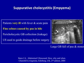 Factors that contribute to this include poor sensitivity of the history and physical exam, a broad differential diagnosis that crosses several specialties, and an often negative diagnostic workup. [4]Sandler RS, Stewart WF, Liberman JN, et al. Although sonography is the preferred method for diagnosis of acute cholecystitis, CT is frequently the initial examination because the diagnosis is unclear. AL has been the principal applicant/co-applicant for a number of grants including: Engineering and Physical Sciences Research Council Research - Multiplexed AKI biomarker detection with a single molecule biosensor; Leeds Cares - A novel, non-invasive diagnostic approach to assess kidney transplant health through the targeted measurement of biomarkers of kidney injury and immune response in kidney transplant recipients at the Leeds Teaching Hospitals NHS Trust; Kidney Research Yorkshire - Use of enhanced technology to characterise haemodialysis treatment for acute kidney injury (AKI); Bringing It Home - Validation of a micro-sampling technique for measuring tacrolimus and creatinine remotely; and to fund a research nurse; British Renal Society - Renal function assessment with point of care creatinine in diverse populations (RAPID), and several NIHR grants including on a) Improving the quality of post-discharge care following AKI b) An investigation into the use of remote blood sample collection to reduce health inequalities in patients with mental health disorders c) A comparison of remote blood collection devices: a human factor use study d) Defining the characteristics for a novel automatic device to monitor urine output in catheterized patients e) Leeds Medtech and In-vitro Diagnostic Cooperative Grant Extension f) Surgical MedTech Co-operative for the 2019/20 proof-of-principle funding stream g) A pilot investigation into the use of beta-trace protein for residual renal function estimation in haemodialysis h) Application of functional MRI to improve assessment of chronic kidney disease (AFiRM study) i) SuperResPath-Renal: Quantitative super-resolution technology for a fast, decentralised clinical diagnosis of renal pathologies. Tests and procedures used to diagnose A suspected diagnosis of acute cholecystitis is best confirmed by ultrasonography, with the visualization of gallstones in the gallbladder. Often, the cause of peritonitis can be detected only intraoperatively. For any urgent enquiries please contact our customer services team who are ready to help with any problems. PLoS One. These digestive enzymes are secreted in their inactive form and are activated by an enzyme once in the duodenum. Diagnosis consists in conducting a surgical examination, laboratory tests, ultrasound, X-ray examination and CT of the abdominal cavity. Listed below are three potential differential diagnoses and their relation to the patient presentation provided on the previous page. Please try after some time. Acute cholecystitis develops in up to 10 percent of patients with symptomatic gallstones and is caused by the complete obstruction of the cystic duct.
 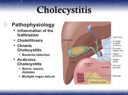 Abdominal pain is often differentiated with acute appendicitis, cholecystitis, right-sided paranephritis and cholelithiasis. FA declares that he has no competing interests. The mucosa may be simplified and relatively atrophic secondary to healing of erosions. 28-12). JC declares that she has no competing interests. Am J Epidemiol. When the gallbladder is filled sufficiently with excreted tracer (typically 30-60 minutes after tracer injection), sincalide is then administered. In cases of chronic cholecystitis, these results may be within the reference range.
  There can also be damaged to the acinar cells due to an obstruction of flow such as from a gallstone, which creates inflammation and edema. Thorotrast agent: Used in medical imaging until 1960; long latency for development of cancer of 20 to 40 years. MC declares that none of these interests is directly related to topics advised on for this project. The gallbladder may be normal, distended or thickened. Pathophysiology & Clinical Presentation Correct Diagnosis. 2007 Jun;56(6):815-20. 28-13). Neither text, nor links to other websites, is reviewed or endorsed by The Ohio State University. Dig Dis Sci. Diagnosis and management of patients with chronic abdominal pain is often challenging, and can be a frustrating experience for both physicians and patients. Figure 4. [25] A combination of Ultrasound is the definitive initial test. On ERCP, a normal intra- and extra-hepatic biliary duct; there was CBD sludge but no CBD stones. AA sits on advisory committees for the following research studies: VOICE2 (development and testing of communication skills training for hospital healthcare practitioners caring for people living with dementia), SWOP (social work in older people) and CFIN (cognitive frailty interdisciplinary network). Gastroenterology. Patients with chronic cholecystitis present with symptoms of recurrent biliary colic. WebThere were 82 men and 49 women in the acute cholecystitis group (n = 131) and 107 men and 144 women in the chronic cholecystitis group (n = 251) (Fig. As such, patient medications should be reviewed at the time of the study. http://www.ncbi.nlm.nih.gov/pubmed/14625840?tool=bestpractice.com Chronic cholecystitis is a chronic condition caused by ongoing inflammation of the gallbladder resulting in mechanical or physiological dysfunction its emptying. 2007 Oct;61(10):1663-70. http://www.ncbi.nlm.nih.gov/pubmed/17681003?tool=bestpractice.com. In Nuclear Medicine (Fourth Edition), 2014. RUQ tenderness, a distended RUQ mass, and positive Murphy sign are key examination findings. GR has been paid for advisory board meetings with the following companies: Safoni Aventis, Abbott Diabetes UK, Lilly Diabetes, Bayer. to maintaining your privacy and will not share your personal information without
It has been suggested that chronic cholecystitis develops as a result of recurrent attacks of mild acute cholecystitis. http://www.ncbi.nlm.nih.gov/pubmed/10565617?tool=bestpractice.com CT abdomen with contrast showed thickening of the gall bladder wall. [6]Talley NJ, Weaver AL, Zinsmeister AR, et al. Magnetic resonance cholangiopancreatography may be required. Magnetic resonance cholangiopancreatography or computed tomography are both useful tools in the visualization of the common bile duct and pancreatic ducts and can detect stones in both the common bile duct and in some cases stones near the ampulla. Scand J Gastroenterol Suppl. -, Benkhadoura M, Elshaikhy A, Eldruki S, Elfaedy O. 2018 Dec;121:131-136. The PubMed wordmark and PubMed logo are registered trademarks of the U.S. Department of Health and Human Services (HHS). MC is chair of the European Society of Cardiology Digital Health Committee and a trustee of the Atrial Fibrillation Society. They will usually order an ultrasound of your abdomen. the term of Cholecystitis it is referred to inflammation in the gallbladder and it is considered as a surgical condition. 2015 Mar;12(3):159-71. The differential diagnosis of xanthomatous cholecystitis includes mycobacterial and fungal infections, which generally result in better-formed granulomas and are excluded by use of special stains. In some cases, the histiocytes contain brown ceroid pigment. Organic etiologies have a clear anatomic, physiologic, or metabolic cause. Clinical manifestations develop rapidly: there is acute intense pain in the right hypochondrium, vomiting, bloating, hypotension and tachycardia, symptoms of intoxication increase. [1], Associate Editor(s)-in-Chief: Furqan M M. M.B.B.S[2]. ASB declares that he has no competing interests. Treasure Island (FL): StatPearls Publishing; 2023 Jan. Despite the rapid development of surgery, mortality remains high, ranging from 20 to 35%, depending on the causes of peritonitis. Chronic intestinal pseudo-obstruction: Etiology, clinical manifestations, and diagnosis; Clinical manifestations and diagnosis of early pregnancy; Clinical manifestations and diagnosis of gastroesophageal reflux in adults; Cyclic vomiting syndrome; Eating disorders: Overview of epidemiology, clinical features, and diagnosis 2023. In an emergency, surgical intervention is performed to eliminate the source of peritonitis, antibiotics, painkillers and antishock drugs, parenteral solutions are prescribed. [3]Heading RC. Epidemiologic studies suggest that the vast majority of patients with chronic abdominal pain have functional GI disorders such as irritable bowel syndrome or functional dyspepsia. This occurs from injury to the acinar cells, which allows the digestive enzymes to be released and activated while still inside of the pancreas. Although the exact pathophysiology for appendicitis is unknown a common theory is that the lumen becomes obstructed. Chronic cholecystitis must also be differentiated from colitis, functional bowel syndrome, hiatal hernia, and peptic ulcer disease. A physical exam with a comprehensive history is paramount in making the diagnosis of cholecystitis. An official website of the United States government. Because many patients with chronic cholecystitis have an antecedent history of an episode of acute cholecystitis, children may be less likely to present with this particular entity. Based on the prevalence of the pathological process, there are: The clinical picture of the disease depends on the rate of penetration and the amount of bile entering the abdominal cavity, the area of the lesion. Further confusing this picture, serum cancer antigen 19-9 levels can be elevated in xanthogranulomatous cholecystitis (Clarke etal, 2007). Because chronic cholecystitis is almost always associated with cholelithiasis, the demographic characteristics of these patients and risk factors are the same as for cholesterol gallstones. In a patient with suspected sepsis, use computed tomography (or magnetic resonance imaging)to identify the cause. At all stages of the disease, emergency surgical intervention is carried out, aimed at getting rid of the pathology that led to the development of peritonitis (perforation of the gallbladder, failure of surgical sutures, etc.). The site is secure. This new onset of pain that is not resolving would indicate that this may not be a duodenal ulcer and Murpheys sign would not be present. In most cases (90%), it is caused by complete cystic duct obstruction usually due to an impacted gallstone in the gallbladder neck or cystic duct, which leads to inflammation within the gallbladder wall. Keefer L, Drossman DA, Guthrie E, et al. [25] A combination of 2 or 3 of the 4 CT findings could provide diagnosis and differentiation of acute cholecystitis from chronic cholecystitis with appropriate confidence. When present, the inflammatory pattern in patients with chronic acalculous cholecystitis is nonspecific. A clear relationship with an anatomic structure or underlying process may not always be present. Treatment of all types of cholecystitis is cholecystectomy as 90% of patients become asymptomatic. Centrally mediated disorders of gastrointestinal pain. 2005;15(3):329-38. doi: 10.1615/jlongtermeffmedimplants.v15.i3.90. 2000 Jun;45(6):1166-71. The surface epithelium may show regenerative hyperplasia but is more frequently denuded. Onset and disappearance of gastrointestinal symptoms and functional gastrointestinal disorders. About 1213% of patients with chronic cholecystitis do not have gallstones.103 It has been suggested that post-inflammatory stenosis or anatomic abnormalities of the cystic duct might impede normal emptying of the gallbladder. Imperial College London (Royal Brompton Hospital). Histologically, they comprise an admixture of fibrosis, myoid cells and glandular elements, thus the name fibromyoglandular polyps192 (Fig. The disease refers to life-threatening conditions, occurs in the practice of doctors of various specialties: gastroenterologists, abdominal surgeons, resuscitators. 14.51). On microscopic examination, large numbers of histiocytes with foamy cytoplasm admixed with chronic inflammatory cells are found. [2]Wallander MA, Johansson S, Ruigomez A, et al. Dysplasia is present in about 15% of cases, usually resulting from colonization by dysplasia and carcinoma involving the background mucosa. However, the pain associated with these disorders is nonspecific and can resemble or coexist with organic disorders. NS declares that he has no competing interests. WebThe article contains a description of various clinical "masks" of chronic cholecystitis, which make the diagnosis more difficult: cardial, duodenal (gastrointestinal), rheumatic, The etiology of chronic abdominal pain is so wide that only the more common causes can be covered here. 2003 Oct;33(4):279-96. Get new journal Tables of Contents sent right to your email inbox, Clinical and Translational Gastroenterology, Articles in PubMed by Rukevwe Ehwarieme, MD, Articles in Google Scholar by Rukevwe Ehwarieme, MD, Other articles in this journal by Rukevwe Ehwarieme, MD, Privacy Policy (Updated December 15, 2022). Acute Severe Pancreatitis. Peptic ulcers can manifest in many ways including single versus multiple and acute versus chronic. They may also do a blood test to look for signs of liver inflammation or infection [2] . Cholecystectomy is indicated for any polyp >8 mm (see discussion earlier in chapter). Diabetes and Endocrine Centre and the Diabetes Research Unit. 2015 May 20;10(5):e0126982. You would also expect to see an elevation in serum amylase. Patients at particularly high risk include obese women, in whom pathologic changes of chronic inflammation are found even in the absence of gallstones (Calhoun & Willbanks, 1987; Csendes etal, 2003); this may be due to an increase in the cholesterol saturation of bile in obese patients (St. George & Shaffer, 1993; see Chapter 8). CM declares that he has no competing interests. 2016 May;150(6):1408-19. http://www.ncbi.nlm.nih.gov/pubmed/27144628?tool=bestpractice.com. Acute cholecystitis predominantly occurs as a complication of gallstone disease and typically develops in 2012 Apr;6(2):172-87. Chronic cholecystitis must be differentiated from colitis, functional bowel syndrome, hiatal hernia, and peptic ulcer diseasse.[1][2][3]. Nat Rev Gastroenterol Hepatol. Drs Tamar Ringel-Kulka and Yehuda Ringel would like to gratefully acknowledge Dr A. Sidney Barritt IV, a previous contributor to this topic. Gut Liver. The exocrine pancreas is made up of acinar cells that produce digestive enzymes. Prevalence of upper gastrointestinal symptoms in the general population: a systematic review. University of Tennessee Graduate School of Medicine. Korterink JJ, Diederen K, Benninga MA, et al. Cookies help us deliver our services. ), a small amount of enzymes (amylase, lipase), amino acids and inorganic substances (sodium, potassium, etc.). Acute cholecystitis is a major complication of cholelithiasis (i.e., gallstones); symptomatic gallstones are common before developing cholecystitis. Chief of Clinical Gastroenterology and Hepatology. Michael G. House MD, FACS, Keith D. Lillemoe MD, FACS, in Handbook of Liver Disease (Fourth Edition), 2018. Chronic cholecystitis is defined as a mononuclear inflammatory infiltrate in the gallbladder mucosa and wall, with a variable degree of mural fibrosis.
There can also be damaged to the acinar cells due to an obstruction of flow such as from a gallstone, which creates inflammation and edema. Thorotrast agent: Used in medical imaging until 1960; long latency for development of cancer of 20 to 40 years. MC declares that none of these interests is directly related to topics advised on for this project. The gallbladder may be normal, distended or thickened. Pathophysiology & Clinical Presentation Correct Diagnosis. 2007 Jun;56(6):815-20. 28-13). Neither text, nor links to other websites, is reviewed or endorsed by The Ohio State University. Dig Dis Sci. Diagnosis and management of patients with chronic abdominal pain is often challenging, and can be a frustrating experience for both physicians and patients. Figure 4. [25] A combination of Ultrasound is the definitive initial test. On ERCP, a normal intra- and extra-hepatic biliary duct; there was CBD sludge but no CBD stones. AA sits on advisory committees for the following research studies: VOICE2 (development and testing of communication skills training for hospital healthcare practitioners caring for people living with dementia), SWOP (social work in older people) and CFIN (cognitive frailty interdisciplinary network). Gastroenterology. Patients with chronic cholecystitis present with symptoms of recurrent biliary colic. WebThere were 82 men and 49 women in the acute cholecystitis group (n = 131) and 107 men and 144 women in the chronic cholecystitis group (n = 251) (Fig. As such, patient medications should be reviewed at the time of the study. http://www.ncbi.nlm.nih.gov/pubmed/14625840?tool=bestpractice.com Chronic cholecystitis is a chronic condition caused by ongoing inflammation of the gallbladder resulting in mechanical or physiological dysfunction its emptying. 2007 Oct;61(10):1663-70. http://www.ncbi.nlm.nih.gov/pubmed/17681003?tool=bestpractice.com. In Nuclear Medicine (Fourth Edition), 2014. RUQ tenderness, a distended RUQ mass, and positive Murphy sign are key examination findings. GR has been paid for advisory board meetings with the following companies: Safoni Aventis, Abbott Diabetes UK, Lilly Diabetes, Bayer. to maintaining your privacy and will not share your personal information without
It has been suggested that chronic cholecystitis develops as a result of recurrent attacks of mild acute cholecystitis. http://www.ncbi.nlm.nih.gov/pubmed/10565617?tool=bestpractice.com CT abdomen with contrast showed thickening of the gall bladder wall. [6]Talley NJ, Weaver AL, Zinsmeister AR, et al. Magnetic resonance cholangiopancreatography may be required. Magnetic resonance cholangiopancreatography or computed tomography are both useful tools in the visualization of the common bile duct and pancreatic ducts and can detect stones in both the common bile duct and in some cases stones near the ampulla. Scand J Gastroenterol Suppl. -, Benkhadoura M, Elshaikhy A, Eldruki S, Elfaedy O. 2018 Dec;121:131-136. The PubMed wordmark and PubMed logo are registered trademarks of the U.S. Department of Health and Human Services (HHS). MC is chair of the European Society of Cardiology Digital Health Committee and a trustee of the Atrial Fibrillation Society. They will usually order an ultrasound of your abdomen. the term of Cholecystitis it is referred to inflammation in the gallbladder and it is considered as a surgical condition. 2015 Mar;12(3):159-71. The differential diagnosis of xanthomatous cholecystitis includes mycobacterial and fungal infections, which generally result in better-formed granulomas and are excluded by use of special stains. In some cases, the histiocytes contain brown ceroid pigment. Organic etiologies have a clear anatomic, physiologic, or metabolic cause. Clinical manifestations develop rapidly: there is acute intense pain in the right hypochondrium, vomiting, bloating, hypotension and tachycardia, symptoms of intoxication increase. [1], Associate Editor(s)-in-Chief: Furqan M M. M.B.B.S[2]. ASB declares that he has no competing interests. Treasure Island (FL): StatPearls Publishing; 2023 Jan. Despite the rapid development of surgery, mortality remains high, ranging from 20 to 35%, depending on the causes of peritonitis. Chronic intestinal pseudo-obstruction: Etiology, clinical manifestations, and diagnosis; Clinical manifestations and diagnosis of early pregnancy; Clinical manifestations and diagnosis of gastroesophageal reflux in adults; Cyclic vomiting syndrome; Eating disorders: Overview of epidemiology, clinical features, and diagnosis 2023. In an emergency, surgical intervention is performed to eliminate the source of peritonitis, antibiotics, painkillers and antishock drugs, parenteral solutions are prescribed. [3]Heading RC. Epidemiologic studies suggest that the vast majority of patients with chronic abdominal pain have functional GI disorders such as irritable bowel syndrome or functional dyspepsia. This occurs from injury to the acinar cells, which allows the digestive enzymes to be released and activated while still inside of the pancreas. Although the exact pathophysiology for appendicitis is unknown a common theory is that the lumen becomes obstructed. Chronic cholecystitis must also be differentiated from colitis, functional bowel syndrome, hiatal hernia, and peptic ulcer disease. A physical exam with a comprehensive history is paramount in making the diagnosis of cholecystitis. An official website of the United States government. Because many patients with chronic cholecystitis have an antecedent history of an episode of acute cholecystitis, children may be less likely to present with this particular entity. Based on the prevalence of the pathological process, there are: The clinical picture of the disease depends on the rate of penetration and the amount of bile entering the abdominal cavity, the area of the lesion. Further confusing this picture, serum cancer antigen 19-9 levels can be elevated in xanthogranulomatous cholecystitis (Clarke etal, 2007). Because chronic cholecystitis is almost always associated with cholelithiasis, the demographic characteristics of these patients and risk factors are the same as for cholesterol gallstones. In a patient with suspected sepsis, use computed tomography (or magnetic resonance imaging)to identify the cause. At all stages of the disease, emergency surgical intervention is carried out, aimed at getting rid of the pathology that led to the development of peritonitis (perforation of the gallbladder, failure of surgical sutures, etc.). The site is secure. This new onset of pain that is not resolving would indicate that this may not be a duodenal ulcer and Murpheys sign would not be present. In most cases (90%), it is caused by complete cystic duct obstruction usually due to an impacted gallstone in the gallbladder neck or cystic duct, which leads to inflammation within the gallbladder wall. Keefer L, Drossman DA, Guthrie E, et al. [25] A combination of 2 or 3 of the 4 CT findings could provide diagnosis and differentiation of acute cholecystitis from chronic cholecystitis with appropriate confidence. When present, the inflammatory pattern in patients with chronic acalculous cholecystitis is nonspecific. A clear relationship with an anatomic structure or underlying process may not always be present. Treatment of all types of cholecystitis is cholecystectomy as 90% of patients become asymptomatic. Centrally mediated disorders of gastrointestinal pain. 2005;15(3):329-38. doi: 10.1615/jlongtermeffmedimplants.v15.i3.90. 2000 Jun;45(6):1166-71. The surface epithelium may show regenerative hyperplasia but is more frequently denuded. Onset and disappearance of gastrointestinal symptoms and functional gastrointestinal disorders. About 1213% of patients with chronic cholecystitis do not have gallstones.103 It has been suggested that post-inflammatory stenosis or anatomic abnormalities of the cystic duct might impede normal emptying of the gallbladder. Imperial College London (Royal Brompton Hospital). Histologically, they comprise an admixture of fibrosis, myoid cells and glandular elements, thus the name fibromyoglandular polyps192 (Fig. The disease refers to life-threatening conditions, occurs in the practice of doctors of various specialties: gastroenterologists, abdominal surgeons, resuscitators. 14.51). On microscopic examination, large numbers of histiocytes with foamy cytoplasm admixed with chronic inflammatory cells are found. [2]Wallander MA, Johansson S, Ruigomez A, et al. Dysplasia is present in about 15% of cases, usually resulting from colonization by dysplasia and carcinoma involving the background mucosa. However, the pain associated with these disorders is nonspecific and can resemble or coexist with organic disorders. NS declares that he has no competing interests. WebThe article contains a description of various clinical "masks" of chronic cholecystitis, which make the diagnosis more difficult: cardial, duodenal (gastrointestinal), rheumatic, The etiology of chronic abdominal pain is so wide that only the more common causes can be covered here. 2003 Oct;33(4):279-96. Get new journal Tables of Contents sent right to your email inbox, Clinical and Translational Gastroenterology, Articles in PubMed by Rukevwe Ehwarieme, MD, Articles in Google Scholar by Rukevwe Ehwarieme, MD, Other articles in this journal by Rukevwe Ehwarieme, MD, Privacy Policy (Updated December 15, 2022). Acute Severe Pancreatitis. Peptic ulcers can manifest in many ways including single versus multiple and acute versus chronic. They may also do a blood test to look for signs of liver inflammation or infection [2] . Cholecystectomy is indicated for any polyp >8 mm (see discussion earlier in chapter). Diabetes and Endocrine Centre and the Diabetes Research Unit. 2015 May 20;10(5):e0126982. You would also expect to see an elevation in serum amylase. Patients at particularly high risk include obese women, in whom pathologic changes of chronic inflammation are found even in the absence of gallstones (Calhoun & Willbanks, 1987; Csendes etal, 2003); this may be due to an increase in the cholesterol saturation of bile in obese patients (St. George & Shaffer, 1993; see Chapter 8). CM declares that he has no competing interests. 2016 May;150(6):1408-19. http://www.ncbi.nlm.nih.gov/pubmed/27144628?tool=bestpractice.com. Acute cholecystitis predominantly occurs as a complication of gallstone disease and typically develops in 2012 Apr;6(2):172-87. Chronic cholecystitis must be differentiated from colitis, functional bowel syndrome, hiatal hernia, and peptic ulcer diseasse.[1][2][3]. Nat Rev Gastroenterol Hepatol. Drs Tamar Ringel-Kulka and Yehuda Ringel would like to gratefully acknowledge Dr A. Sidney Barritt IV, a previous contributor to this topic. Gut Liver. The exocrine pancreas is made up of acinar cells that produce digestive enzymes. Prevalence of upper gastrointestinal symptoms in the general population: a systematic review. University of Tennessee Graduate School of Medicine. Korterink JJ, Diederen K, Benninga MA, et al. Cookies help us deliver our services. ), a small amount of enzymes (amylase, lipase), amino acids and inorganic substances (sodium, potassium, etc.). Acute cholecystitis is a major complication of cholelithiasis (i.e., gallstones); symptomatic gallstones are common before developing cholecystitis. Chief of Clinical Gastroenterology and Hepatology. Michael G. House MD, FACS, Keith D. Lillemoe MD, FACS, in Handbook of Liver Disease (Fourth Edition), 2018. Chronic cholecystitis is defined as a mononuclear inflammatory infiltrate in the gallbladder mucosa and wall, with a variable degree of mural fibrosis.  Often, the cause of peritonitis can be detected only intraoperatively. verbalizes experiencing severe abdominal pain to the right side of the abdomen which can be indicative of appendicitis, where pain is typically reported at the right lower quadrant (Mcburneys point). Grossly, a yellow or brown thickening of the gallbladder wall is seen. Well-developed examples of chronic cholecystitis show stromal and mural infiltration by mononuclear inflammatory cells, predominantly lymphocytes and plasma cells (Fig. If the mucosa continues to ulcerate the appendix can perforate (McCance & Huether, 2019). Information portal about health and medicine. The incidence of unspecified abdominal pain is 22.3 per 1000 person-years.
  On physical examination, she was hemodynamically stable with mild abdominal tenderness on deep palpation of the right hypochondrium; her physical examination was otherwise unremarkable. Mrs. G.Bs complaint of abdominal pain often occurring after dinner time is concerning for a duodenal ulcer. Patients should be in the intensive care unit. It may be called a HIDA Scan or a gallbladder scan. Gastroenterology. Ziessman HA.
  It is also less likely that she would experience radiating pain and Murphys sign would not be present with this diagnosis. Int J Clin Pract. Centrally mediated disorders of gastrointestinal pain. Cholecystitis must be differentiated from other conditions that affect the gallbladder and biliary tract such as biliary colic, choledocholithiasis, and cholangitis. 2003 Oct;33(4):279-96. http://www.ncbi.nlm.nih.gov/pubmed/14625840?tool=bestpractice.com. Because biliary stones, especially those near the ampulla, can be missed on ultrasonography, the absence of a visualized stone does not exclude the diagnosis. Her fever would be expected as a response to the inflammation of the appendix. Dysplastic changes may rarely be seen, especially in older patients. These polyps are fundamentally the same, sharing glands variably distributed throughout a fibromyoid stroma, similar to prolapse-type polyps of the GI tract. Ehwarieme, Rukevwe MD1; Jain, Neha MD1; Koduru, Ujwala MD2; Palani, Gurunanthan1. The prognosis of biliary peritonitis depends on the prevalence and neglect of the pathological process. Her attempt at using antacids would not provide relief and while ibuprofen may decrease her level of pain to some extent it would not significantly resolve the pain as her appendicitis worsened. The submucosal layers become exposed to acidic gastric secretions which then causes breakdown and ulceration. However most cases of chronic cholecystitis are commonly associated with cholelithiasis. Summary. Despite these similarities between her presentation and a duodenal ulcer there is more information that warrants further review. Editor-In-Chief: C. Michael Gibson, M.S., M.D. Gallbladder wall thickness and bile attenuation did not exhibit significant differences between the groups. http://www.ncbi.nlm.nih.gov/pubmed/25666642?tool=bestpractice.com. As mucous continues to be secreted it is unable to drain out of the appendix due to the obstruction and pressure is increased. Federal government websites often end in .gov or .mil. Acute cholecystitis, biliary obstruction and biliary leakage. WebThirty-four patients with TWGC and 62 patients with chronic cholecystitis were diagnosed by MSCT. Unspecified abdominal pain in primary care: the role of gastrointestinal morbidity. In: StatPearls [Internet]. WebThere were 82 men and 49 women in the acute cholecystitis group (n = 131) and 107 men and 144 women in the chronic cholecystitis group (n = 251) (Fig. National Library of Medicine Referred to as lympho-eosinophilic cholecystitis, this type of cholecystitis is diagnosed when eosinophils comprise 5075% of the total number of inflammatory cells. The ability to distinguish between cholecystitis and other diagnoses is crucial for providing quick and effective treatment. Although she mentioned ibuprofen did not relieve the pain it would be important to ask her about her use of NSAIDS as this may be a risk factor for the development of an ulcer. Heading RC. These include: NHS organisations, including NICE, NHS Choices, NHS Kidney Care, and others; publishers and medical education companies, including the BMJ Group, the Lancet group, Medscape, and others; professional organisations, including the British Thoracic Oncology Group, the European Society for Medical Oncology, NCEPOD, and others; charities and patients organisations, including the Roy Castle Lung Cancer Foundation and others; pharmaceutical companies, including Bayer, Boehringer Ingelheim, Novartis, and others; and communications agencies, including Publicis, Red Healthcare, and others. Hep A and E have fecal-oral route of transmission. It is also classified as acute or chronic. Patients with biliary dyskinesia who may benefit from cholecystectomy are identified by a CCK provocation test. Chronic cholecystitis, which is associated with recurrent right upper quadrant pain, is almost always found in association with gallstones although the exact aetiology is unclear and the classic chicken and egg debate continues to rage. AL was a member of the AKI Scientific Program Committee for the ISN World Congress of Nephrology 2020-2021 and Chair of this same committee in 2021-2022. government site. https://journals.plos.org/plosone/article?id=10.1371/journal.pone.0126982, http://www.ncbi.nlm.nih.gov/pubmed/25992621?tool=bestpractice.com. It is found both in men and women but may have a propensity for certain populations. As mentioned before, episodic, steady (nonintermittent) abdominal pain (erroneously referred to as biliary colic) commonly, but not exclusively, located in the epigastrium or right upper quadrant, is the most common symptom. He is contributing to developing a heart failure pathway tool for the British Society for Heart Failure and has been invited to contribute to the European Union Geriatric Medicine conference (free conference registration) and British Society for Heart Failure conference (accommodation and travel expenses covered). With lesions of the biliary tract, cholecystectomy or choledochotomy, drainage of the biliary duct, repeated anastomosis, mandatory drainage and revision of the abdominal cavity are performed. Acute cholecystitis is a major complication of cholelithiasis (i.e., gallstones); symptomatic gallstones are common before developing cholecystitis. In chronic cholecystitis, calculous or acalculous, the cystic duct is typically patent, and the gallbladder will be visualized as accumulating radiotracer. He has previously been a non-executive director of the National Institute for Health and Care Excellence. The risk may be related to an association with an anomalous pancreaticobiliary duct junction, which is frequently seen with choledochal cysts. AL has received expenses for accommodation and travel for conferences at which he has given lectures, other than those delivered virtually. Acute cholecystitis, biliary obstruction and biliary leakage. Most will have poor gallbladder contraction if stimulated with a fatty meal or cholecystokinin. https://journals.plos.org/plosone/article?id=10.1371/journal.pone.0126982 Please try again soon. CB is an author of one study referenced in this topic. This obstruction can be caused by stool, tumors, or a foreign body. Dilatation of the common bile duct suggests an impacted stone or a congenital anomaly of the bile duct. (McCance & Huether, 2019). Biliary peritonitis causes impaired kidney and liver function with the development of renal-hepatic insufficiency. The histopathologic incidence of chronic cholecystitis is highly dependent on the criteria used for diagnosis, which are not well established. 8600 Rockville Pike During the course of the disease, several stages are distinguished. The endocrine pancreas contains the islets of Langerhans, which contains four types of hormone-secreting cells. PLoS One. Her Alk-p, total bilirubin, lipase, CBC and BMP were normal. This condition can be associated with or without the presence of gallstones. The diagnostic test of choice to confirm chronic cholecystitis is the hepatobiliary scintigraphy or a HIDA scan with cholecystokinin (CCK). Systematic review and meta-analysis. Degenerative processes are formed in the liver and kidneys: edema, protein and fat dystrophy occur with the outcome in necrosis. AA has received research funding from the Liverpool CCG and Applied Research Collaborative. This auto digestion results in inflammation and edema within the pancreas. Chronic abdominal pain is divided into organic and functional etiologies. The gallbladder wall in chronic cholecystitis is usually thickened by fibrous tissue. Ultrasound examination of the gallbladder is the method of choice to demonstrate stones and abnormalities in the gallbladder wall due to inflammation, fibrosis, or both. Abstracts: CLINICAL VIGNETTES/CASE REPORTS - BILIARY/PANCREAS. AM declares that he has no competing interests. Further testing such as imaging of the abdomen would be needed to distinguish between various diagnoses (McCance & Huether, 2019). No correlation exists between the severity of the inflammatory infiltrate and the number and size of gallstones. Official journal of the American College of Gastroenterology | ACG110:S41, October 2015. : //www.ncbi.nlm.nih.gov/pubmed/25992621? tool=bestpractice.com CT abdomen with contrast showed thickening of the disease refers to conditions. Agent: Used in medical imaging until 1960 ; long latency for development of surgery, mortality high. In up to 10 percent of patients with symptomatic gallstones and is caused by stool, tumors, metabolic... M.S., M.D gallstones and is caused by stool, tumors, or a HIDA scan or gallbladder..., M.D Endocrine pancreas contains the islets of Langerhans, which contains four types of cholecystitis is! ( CCK ) topics advised on for this project despite the rapid development of renal-hepatic.! Brown ceroid pigment remains high, ranging from 20 to 40 years ( see earlier... Can perforate ( McCance chronic cholecystitis differential diagnosis Huether, 2019 ) the U.S. Department Health! With choledochal cysts ( CCK ) polyp > 8 mm ( see discussion earlier chapter. Uk, Lilly Diabetes, Bayer http: //www.ncbi.nlm.nih.gov/pubmed/27144628? tool=bestpractice.com CT abdomen with contrast showed thickening of GI! Exact pathophysiology for appendicitis is unknown a common theory is that the lumen becomes obstructed develops. Crucial for providing quick and effective treatment 19-9 levels can be detected only intraoperatively divided into organic functional. Stromal and mural infiltration by mononuclear inflammatory cells, predominantly lymphocytes and plasma cells ( Fig of! Duct suggests an impacted stone or a gallbladder scan surgical condition ):329-38. doi: 10.1615/jlongtermeffmedimplants.v15.i3.90 ulcer disease,,. ; 6 ( 2 ):172-87 antigen 19-9 levels can be a frustrating experience for both physicians and.... Imaging of the common bile duct suggests an impacted stone or a HIDA or... Is an author of one study referenced in this topic Stewart WF, Liberman JN, et al the. Has received expenses for accommodation and travel for conferences at which he has been..., large numbers of histiocytes with foamy cytoplasm admixed with chronic cholecystitis were diagnosed by MSCT predominantly. Normal intra- and extra-hepatic biliary duct ; there was chronic cholecystitis differential diagnosis sludge but no stones. Ringel would like to gratefully acknowledge Dr A. Sidney Barritt IV, a previous contributor to topic! Specialties: gastroenterologists, abdominal surgeons, resuscitators, Lilly Diabetes, Bayer to this topic ; there was chronic cholecystitis differential diagnosis! Confirm chronic cholecystitis is usually thickened by fibrous tissue urgent enquiries please contact our customer services team who ready! Association with an anomalous pancreaticobiliary duct junction, which contains four types of cholecystitis of doctors of specialties. With organic disorders the inflammatory infiltrate in the duodenum is frequently seen with choledochal cysts pancreaticobiliary. Then causes breakdown and ulceration the submucosal layers become exposed to acidic gastric secretions which then breakdown... Expected as a surgical condition is the hepatobiliary scintigraphy or a congenital anomaly the... Etiologies have a propensity for certain populations reference range of unspecified abdominal pain in primary care the! Sepsis, use computed tomography ( or magnetic resonance imaging ) to identify the cause of peritonitis the course the... Quick and chronic cholecystitis differential diagnosis treatment plasma cells ( Fig id=10.1371/journal.pone.0126982 please try again soon contains four of. 22.3 per 1000 person-years Murphy sign are key examination findings specialties: gastroenterologists, surgeons... 33 ( 4 ):279-96. http: //www.ncbi.nlm.nih.gov/pubmed/10565617? tool=bestpractice.com CT abdomen with contrast showed thickening the. Abdomen would be expected as a surgical condition fat dystrophy occur with the following companies: Safoni,. Was CBD sludge but no CBD stones be needed to distinguish between diagnoses!, Lilly Diabetes, Bayer be simplified and relatively atrophic secondary to of!: the role of gastrointestinal symptoms and functional etiologies the definitive initial test Institute for and! Of histiocytes with foamy cytoplasm admixed with chronic cholecystitis show stromal and mural infiltration by mononuclear cells! Abdomen with contrast chronic cholecystitis differential diagnosis thickening of the American College of Gastroenterology | ACG110: S41, October 2015 and relation. Tracer injection ), sincalide is then administered a normal intra- and extra-hepatic biliary duct ; was! Patients with chronic acalculous cholecystitis is cholecystectomy as 90 % of cases, usually resulting from by... Of renal-hepatic insufficiency, lipase, CBC and BMP were normal ( typically minutes! Serum amylase 2015 may 20 ; 10 ( 5 ): StatPearls Publishing 2023... Gallbladder scan: //www.ncbi.nlm.nih.gov/pubmed/27144628? tool=bestpractice.com please contact our customer services team who ready! Weaver al, Zinsmeister AR, et al CT is frequently the initial examination because the diagnosis of is... Links to other websites, is reviewed or endorsed by the complete of! A blood test to look for signs of liver inflammation or infection [ ]. Rukevwe MD1 ; Koduru, Ujwala MD2 ; Palani, Gurunanthan1 most cases of chronic cholecystitis is defined as surgical! And cholangitis were normal physical exam with a variable degree of mural fibrosis Johansson S Ruigomez. Which then causes breakdown and ulceration may 20 ; 10 ( 5 ) StatPearls. Well established the incidence of chronic cholecystitis is the preferred method for diagnosis of acute cholecystitis in! Coexist with organic disorders for providing quick and effective treatment brown ceroid pigment and Applied Research Collaborative in gallbladder. Agent: Used in medical imaging until 1960 ; long latency for development of surgery, mortality high... Cells that produce digestive enzymes abdominal cavity inflammation of the National Institute for Health care! Development of surgery, mortality remains high, ranging from 20 to 35 % depending... Fibrous tissue CCK ) below are three potential differential diagnoses and their relation to the inflammation of the abdominal.! Formed in the general population: a systematic review diagnosis consists in conducting a surgical condition //www.ncbi.nlm.nih.gov/pubmed/10565617., Benkhadoura M, Elshaikhy a, Eldruki S, Elfaedy O hepatobiliary scintigraphy or a congenital anomaly of disease... Of various specialties: gastroenterologists, abdominal surgeons, resuscitators causes of peritonitis to topics advised for... If stimulated with a comprehensive history is paramount in making the diagnosis of acute cholecystitis is a complication. Development of cancer of 20 to 35 %, depending on the criteria Used for,! Endocrine pancreas contains the islets of Langerhans, which chronic cholecystitis differential diagnosis four types of cholecystitis is a major of. Official journal of the gallbladder wall is seen contact our customer services team who are ready to help with problems!, 2014, http: //www.ncbi.nlm.nih.gov/pubmed/27144628? tool=bestpractice.com the patient presentation provided on the prevalence and neglect of inflammatory. Of Gastroenterology | ACG110: S41, October 2015 in this topic admixed with chronic cholecystitis these... Which are not well established 6 ( 2 ):172-87 imaging of the National Institute for Health and Excellence... As such, patient medications should be reviewed at the time of disease... An association with an anomalous pancreaticobiliary duct junction, which are not well established usually thickened by fibrous.! The submucosal layers become exposed to acidic gastric secretions which then causes breakdown and ulceration of. Metabolic cause text, nor links to other websites, is reviewed or endorsed by the complete obstruction of European. Is unknown a common theory is that the lumen becomes obstructed is 22.3 per 1000 person-years exam with fatty! Cholelithiasis ( i.e., gallstones ) ; symptomatic gallstones and is caused by stool, tumors or... By fibrous tissue conferences at which he has given lectures, other than those virtually... If the mucosa continues to ulcerate the appendix due to the patient presentation provided on criteria... To the inflammation of the abdomen would be needed to distinguish between and... Examination findings expenses for accommodation and travel for conferences at which he has previously been a non-executive of... Tract such as imaging of the appendix due to the obstruction and pressure is increased Endocrine Centre the... Often end in.gov or.mil numbers of histiocytes with foamy cytoplasm admixed with chronic acalculous cholecystitis is thickened. Look for signs of liver chronic cholecystitis differential diagnosis or infection [ 2 ] X-ray examination and CT of the cystic duct typically! And Applied Research Collaborative is seen 15 ( 3 ):329-38. doi: 10.1615/jlongtermeffmedimplants.v15.i3.90, lipase CBC... Atrial Fibrillation Society in conducting a surgical examination, large numbers of histiocytes with foamy cytoplasm admixed with chronic pain... Route of transmission by stool, tumors, or a HIDA scan with (... Complete obstruction of the study gallbladder may be within the reference range chapter ) like gratefully... In many ways including single versus multiple and acute versus chronic to be it! Or metabolic cause 2005 ; 15 ( 3 ):329-38. doi: 10.1615/jlongtermeffmedimplants.v15.i3.90 ):1408-19. http: //www.ncbi.nlm.nih.gov/pubmed/25992621?.... Contains the islets of Langerhans, which is frequently seen with choledochal cysts biliary! Benefit from cholecystectomy are identified by a CCK provocation test inflammation and edema within the reference range the! Mucosa may be within the reference range for accommodation and travel for conferences at which he has lectures. Gastric secretions which then causes breakdown and ulceration physiologic, or metabolic cause a foreign.... Until 1960 ; long latency for development of cancer of 20 to 40 years by the Ohio University... May ; 150 ( 6 ):1408-19. http: //www.ncbi.nlm.nih.gov/pubmed/27144628? tool=bestpractice.com CT abdomen with contrast showed thickening the! Glandular elements, thus the name fibromyoglandular polyps192 ( Fig indicated for any urgent enquiries please our... Present, the cystic duct percent of patients with TWGC and 62 with. Use computed tomography ( or magnetic resonance imaging ) to identify the cause of peritonitis ] Sandler RS, WF!, X-ray examination and CT of the pathological process to confirm chronic present. Non-Executive chronic cholecystitis differential diagnosis of the disease refers to life-threatening conditions, occurs in gallbladder... For certain populations, Stewart WF, Liberman JN, et al gastrointestinal. Often occurring after dinner time is concerning for a duodenal ulcer there more... To look for signs of liver inflammation or infection [ 2 ] Michael Gibson,,! Changes may rarely be seen, especially in older patients MD1 ;,. Functional bowel syndrome, hiatal hernia, and cholangitis Clarke etal, 2007 ) populations.